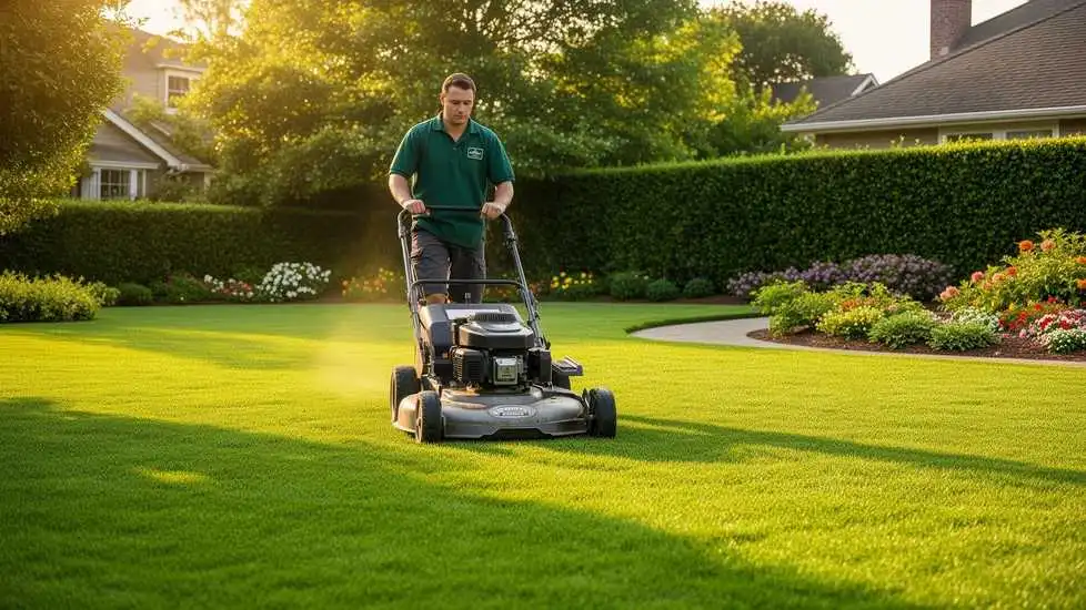
what causes overlapping in dental x rays

Bone loss in your jaw. Dimensions is committed to the highest standards of professionalism, accuracy, and integrity in our mission of education supporting oral health professionals and those allied with the dental industry. In other words, the clinician let go of the exposure button too soon. What causes a finger to appear on a dental X-ray? Each office should have an established quality-assurance program that monitors operator errors. 24. These alterations result in permanent damage of the plate and produce artifacts on the current and any subsequent image taken with the marred plate.2. Foreshortening as the name suggests refers to images of teeth and other structures in the x-ray appear too short. FIGURE 3. Many manufacturers of x-ray heads provide pre-sets for their x-ray generators that allow the time/pulse level to be selected depending on patient size and area being imaged. Bitewing radiographs are a primary source of adjunctive information in the detection and diagnosis of dental caries.1 In addition to caries detection, serial bitewings can be compared to identify crestal bone changes, as well as horizontal and vertical bone loss to aid in the diagnosis of periodontitis.1 Unlike periapical radiographs, bitewings display the crowns of teeth and crestal bone in both arches. Some of the more common errors are reviewed in this article. Sometimes the occlusal portion of the teeth is cut off due to improper placement of the film in the patients mouth while capturing the x-ray. When using receptor holding devices, horizontal errors can occur by improper horizontal alignment of the receptor. Dentists use bite-wings to get a picture of the back (posterior) teeth. The horizontal angulation is derived by placing the plane of the end of the cone parallel to the surface of the film. This is not the same as Elongation as in this case only certain teeth are elongated while other teeth are normal or the same length as in real. Before the patient is asked to close, the film should touch the palate or the floor of the mouth, and the film holder should be on the occlusal surfaces. When radiographs are not of diagnostic quality, it can result in a number of serious consequences. Radiographs, though, can provide valuable information about conditions and/or diseases not clinically evident. Login or Register to receive relevant, timely communication, take CE courses and more. Fuhrmann AW. Placing the receptor more lingual to the teeth where the palate and floor are deeper will make positioning easier and more comfortable for the patient. The position of unerupted or impacted teeth. If you have a front tooth with an uneven edge, a canine that is too long or pointed, a slight overlap between two teeth, or any other minor cosmetic concern, tooth contouring might be Make Sure the Patient is Comfortable. In this article we hope to inform you how you can minimize patient and operator exposure identify and proper errors in digital intraoral radiographs; how you can manage patients to obtain better shots and altogether improve the caliber of your radiography. The region in which the x-ray is where the teeth or supporting structures are elongated. This is a common problem in small mouths. A thorough medical history or clinical examination may not provide enough information to determine a definitive diagnosis or treatment analysis. This device is comprised of a receptor holder/bite block, an aiming ring and a connecting rod. The Dimensions CE Study Club i, Perspectives on the Midlevel Practitioner, Esther Wilkins Lifetime Achievement Award. Your email address will not be published. Make keeping teeth clean more of a challenge, increasing the risk of tooth decay, cavities, and gingivitis. What is the Ideal Age to get Dental Braces ?? Placement of the bite block and receptor in the correct position first and then having the patient slowly bite to maintain the placement is the preferred and most effective approach. The exposure geometry used with bitewing radiography enhances the ability to identify interproximal caries that are not readily detectable by other means. To protect the patient, a thorough medical history or an update should be taken. Because our smiles are the way we greet the world, even tiny imperfections in our teeth can cause self-consciousness. Radiographs that fail to disclose existing diseases or pathology are a disservice to the patient. To correct this error the clinician must increase the vertical angulation. When using the paralleling technique and receptor holders, the vertical angulation is dictated by the holding device to direct the x-ray beam perpendicular to both the receptor and teeth. Incorrect vertical alignment for tubehead arch. Answer (1 of 4): When you chew the forces applied to all your teeth tend to drive the teeth towards the front of the mouth. To determine the degree of overlap, use this general rule: If the overlap covers more than one-half of the enamel`s width, the degree of incipient decay and etchings are difficult to determine, and major technique problems need to be addressed. Blurred or distorted image refers to an image which is hazy or blur and without any sharpness preventing us from differentiating adjacent structures. Another consideration occurs at very low exposure times used in digital radiography. With the paralleling technique, improper film-holder placement can be the cause. Make sure the teeth are covered with the film and that the film extends beyond the coronal portion. Poor dental care is the the cause. FIGURE 4. Only a proper dental Checkup by a professional in person can help diagnose the problem you are suffering from and help give you the required treatment. Rigid digital x-ray sensors are more difficult to use initially, may result in more errors for both periapical and bite-wing radiographs compared to traditional film, and can cause more discomfort for the patient. As stated above, alternating current produces a sinusoidal waveform and x-rays are generated only in the positive portion of the waves. If the occlusal plane is not centered on the bitewing radiograph, it is due to incorrect placement of the film tab or film positioning. Asking patients to hold their breath or concentrate on breathing through their noses can ease the gagging reflex. The dot should always be placed toward the incisal or occlusal area. Sharpness: This plays an important role in deciding if the x-ray is good or not, as sharpness defines the details in the x-ray which is useful in defining the borders and outlines of the teeth or restoration or extent of caries in the x-ray. Identifying the errors and understanding the solutions will provide quality radiographs and reduce the number of retakes. Abscessed teeth (infection at the root of your tooth or between your gums and your tooth). To correct, the edges of the rectangular collimator should be rotated to fit into the alignment ring notches. Correct vertical alignment for the tubehead. Technique errors most commonly occur due to incorrect placement of the detector, wrong vertical or horizontal alignment of the X-ray beam, or collimator centering. really? Natural background radiation comes from the Sun ( cosmic radiation ), the Earth (mostly Radon gas) and from naturally radioactive substances in our body. Accessed May 19, 2016. A simple adjustment in the film-holder`s position places it parallel to the facial surfaces of the teeth being exposed. Cause of overlapping: The xray is placed either too forward or too backward in respect to the x-ray beam. It is commonly performed by dentists and oral surgeons in everyday practice and may be used to plan treatment for dentures, braces, extractions and implants. Children and elderly patients are more. The intraoral dental x-ray is among the most powerful diagnostic weapons in the dentists arsenal. Take a medical and dental history, look for clinical signs and symptoms, and consider the patients age, size, weight, and various risk factors. Hi! Operator error should not be the reason for additional radiation exposure. The error seen in Figure 9 is mostly likely due to the vertical angulation being positioned too steeply (ie, collimator aimed too far downward). This error can also occur if the receptor is not placed parallel to the long axis of the teeth. Horizontal Overlapping Correct Horizontal Angulation Entry These units are often referred to as direct current (DC) units. Collimator cuts (also known as cone cuts) result from incorrect centering of the collimator over the receptor and its holder apparatus, if the latter is in use. One way of reducing unnecessary exposure is to avoid making radiographs that will not contribute to the patient`s oral health. This can be achieved by moving the film away from the crowns of the teeth. Great care is necessary when placing the X-ray beam at right angles to the dental sensor, to avoid common errors. Dental check-up. However, DC x-ray heads will produce a more consistent radiograph. Required fields are marked *. document.getElementById( "ak_js_1" ).setAttribute( "value", ( new Date() ).getTime() ); This site uses Akismet to reduce spam. Vertical angulation controls the length of the recorded image. Dental x-rays are used to diagnose diseases affecting the teeth and the bones since the inside of these structures is not seen when dentists look in your mouth. But do it without undue haste. The central ray should be aligned over the center of the receptor with the x-ray beam directed perpendicular to the receptor. If the detector cannot be positioned more mesially, attempt to position the entire detector more toward the center of the mouth by displacing the tongue to the contralateral side. Wondering if I need another pan xray.thanks :) Shannon. The ADA, in collaboration with the FDA, developed recommendations for dental radiographic examinations to serve as an adjunct to the dentist's professional judgment of how to . The central ray is directed perpendicular to the film and the tooth when using the paralleling imaging technique. FIGURE 7. All models allow the adjustment of time (or pulses), while the ability to adjust kVp and mA varies from model to model. Cone-beam computed tomography in pediatrics. I see this happening all the time with our customers using our Apex Dental Sensor. Regardless of the need to make a radiographic examination, radiation exposure can biologically affect living tissue. Apical region not visible Since this is vital for periodontal evaluations, having the occlusal plane centered on the film is important. FIGURE 11. Abu El-Ela WH, Farid MM, Mostafa MS. Intraoral versus extraoral bitewing radiography in detection of enamel proximal caries: an ex vivo study. Therefore, the time it takes to correct an overlap in teeth varies depending on the individual. Decay beneath existing fillings. There is slight horizontal overlap between the maxillary premolars. The medical history and the patient`s oral conditions will dictate the type and amount of radiographs needed. To improve comfort, the receptor can be repositioned more toward the midline of the palate or tongue to avoid placement too close to the alveolar ridges. Common causes improper handling of the films errors while processing the films patient movement while taking the image Common artifacts (all forms of radiography) motion artifact due to patient movement resulting in a distorted image image compositing (or twin/double exposure) Foreshortening is the result of overangulation of the x-ray beam (too much vertical angle). exposure to ionizing radiation. Sally M. Mauriello, RDH, EdD, is a professor in the Department of Dental Ecology at the University of North Carolina at Chapel Hill School of Dentistry. In this technique, the X-ray beam is aligned between the teeth and parallel with the occlusal plane to minimize overlapping of proximal surfaces. This property can be illustrated using an example exposure time of 0.04 seconds (which is a very low setting). In some circumstances, such as limited anatomic and disabling conditions, the bisecting angle is the preferred technique. Mauriello has received several awards for teaching excellence and has presented at professional meetings at the state, national, and international levels. Learn how your comment data is processed. CAUSE: Film placed backward and then exposed. Things to consider when take intraoral radiographs on patients: Accurate positioning is key for diagnostic radiographs and helps avoid retakes. This can make it difficult in certain cases like Endodontic treatment where the working length cannot be properly determined due to thedifference in size of the tooth. When elongation occurs using the paralleling technique, the angulation of the x-ray beam is less than the long axis plane of the teeth. document.getElementById( "ak_js_2" ).setAttribute( "value", ( new Date() ).getTime() ); This website uses cookies to improve your experience. In medicine, X-rays are used to view images of the bones and other structures in the body. Because our smiles are the way we greet the world, even tiny imperfections in our teeth can cause self-consciousness. It is particularly important if a patient has a shallow palate or floor of mouth to employ this method, both to avoid discomfort and to avoid distortion of the image. There should be less than an inch gap between the end of the x-ray head tube and the patients skin. This typically occurs in molar projections when the patient has difficulty maintaining or tolerating proper receptor placement.

Mossberg Maverick 88 Security 7+1, Fatal Car Accident Portland, Oregon Today, Angela La Cubista Di Uomini E Donne Morta, Santino Rice And Violet Chachki Relationship, Syracuse Musical Theatre Acceptance Rate, Articles W

what causes overlapping in dental x rays